Hearing Tests
Types of hearing tests
Audiogram
The basic hearing test or audiogram tests one's ability to hear pure tones in each ear. Best results are obtained by a trained audiologist in a special soundproof testing booth. Simple tests, such as ones done in many schools, may be useful for screening, but a careful audiogram is necessary for accurate diagnosis of most hearing problems.
A complete audiogram will test both the bone conduction (the ability to hear a sound when it transmitted through bone) and the air conduction (the ability to hear a sound when it transmitted through air). A comparison between these two type of conduction can be very useful in localizing which part of the hearing mechanism is responsible for the loss. In particular, the test is useful in determining if the loss is due to problems with the portion of the middle ear that conducts sound from the ear canal to the inner ear (in which case it would be called a "conductive" hearing loss) or if it is due to the inner ear or the nerve that conducts the sound signals to the brain (in which case it would be called a "sensorineural" hearing loss).
The results of audiograms are most often displayed in graph form. This graph shows the amount of hearing loss expressed in units called decibels at different sound frequencies (also called Hertz). High frequencies correspond to high tones, and low frequencies are low tones. Most audiograms go from around 250 hertz to 4000 hertz. A loss up to 20 decibels on this graph is considered "normal." Hearing losses over 20 decibels are considered abnormal.
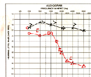
In the audiogram below, the red tracings are for the right ear, and the black tracings are for the left. The round circles represent the air conduction and the brackets are the bone conduction. Low frequencies are to the left and high frequencies are on the right. A normal audiogram would show all hearing loss of 20 dB or less. (The measurements would all be above 20 dB. This audiogram shows a normal tracing on the left, and a sensorineural (nerve) hearing loss on the right.
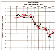
In this audiogram there is a sensorineural hearing loss on both sides in the high frequencies.
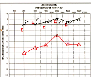
In this audiogram the left side is normal. On the right side the bone conduction (the "[" symbol) is normal for all frequencies, but the air conduction on the right is decreased. This is called an "air-bone" gap and implies a problem with the conductive portion of hearing. It could be caused by a problem with the tympanic membrane or the bones of the middle ear.
The tympanogram is a test that measures how easily the eardrum vibrates back and forth and at what pressure is the maximum. The middle ear is normally filled with air at a pressure equal to the surrounding atmosphere. If the middle ear is filled with fluid, the eardrum will not vibrate properly and the tympanogram will be flat. If the middle ear is filled with air but at a higher or lower pressure than the surrounding atmosphere, the tympangram will be shifted in its position.
The tympanogram is a quick and easy test. A special probe is placed up against the ear canal, like an ear plug, then the equipment automatically makes the measurements.
Auditory Brainstem Response (ABR)
The ABR is a special hearing test that can be used to track the nerve signals arising in the inner ear as they travel through the hearing nerve (called the auditory nerve) to the region of the brain responsible for hearing. The test is useful because it can tell us where along that path the hearing loss has occurred. For example, the ABR is often used for individuals with a sensironeural (nerve) loss in just one ear. This loss can sometimes be caused by a benign (non-cancerous) tumor on the auditory nerve. If the ABR is normal along that region of the path, the chances of having this tumor are quite small.
The ABR can also be used on small infants since it requires no conscious response from the person being tested. A small speaker is placed near the ear which produces clicking sound. Special electrodes automatically record the nerve signal; the patient can even be asleep during the test.
Videonystagmography (VNG)
The VNG is not a hearing test but rather a special test of the balance mechanism of the inner ear. The test has 3 main parts:
- Ocular mobility testing entails following a moving dot with your eyes to look for any slowness or inaccuracies in your ability to follow visual targets.
- Positional testing looks at eye movements during different positions of moving the head and body.
- Caloric testing involves stimulating both ears with warm and then cool air. This change in temperature stimulates the inner ear which in turn causes reflex movements of the eyes.
From the 3 main parts of VNG, eye movements are recorded and measured. Analyzing these eye movements reveals how well the balance mechanism is functioning and may indicate if a central or neurological problem or problem in the inner ear exists.
- Acoustic Neuroma
- Bone-Anchored Hearing Aids
- Cochlear Implants
- Chronic Ear Infections
- Do I Really Need Two Hearing Aids?
- Ear Tubes
- Facial Nerve Weakness
- How Hearing Works
- Hearing Tests
- Tips for Better Hearing
- Meniere's Disease
- Otosclerosis
- Perforations of the Eardrum
- Tinnitus
- Types of Hearing Impairment
- Hearing Aids