About Laryngeal Cancer
Common questions about laryngeal cancer
Laryngeal cancer is the term given to a malignant tumor of the larynx, or voice box. Laryngeal cancer sometimes is simply called throat cancer, though the term is somewhat misleading since the throat includes regions other than just the larynx.
The most common type of laryngeal cancer is known as squamous cell carcinoma (SCCA). The lining of a portion of the larynx is made up of a type of cell called squamous cells. When a malignant tumor arises in these cells, it is called laryngeal carcinoma or laryngeal cancer.
What causes laryngeal cancer?
Laryngeal cancer is most often caused by a long exposure to tobacco and to alcohol. Its incidence is much higher in people who have a history of heavy smoking or drinking. It can occur in people who have never smoked or who never drank alcohol, but this is very rare.
What are some symptoms of laryngeal cancer?
Typical symptoms of laryngeal cancer are throat pain, hoarseness, ear pain, breathing difficulty, or coughing up blood. A lump in the neck could also be a sign of a tumor in the head and neck region. Though these symptoms may be caused by other less serious illnesses, if they occur they should be brought to a physician's attention right away.
How is laryngeal cancer diagnosed?
Laryngeal cancer is diagnosed by a careful examination by a physician who specializes in throat disorders. The first step is a complete patient history and physical examination. In order to look at the larynx, the physician must either use a mirror or a flexible fiberoptic scope that passes through the nose or the mouth into the back of the throat.
If the physician sees a suspicious mass, the next step is a biopsy. The procedure is called laryngoscopy and is usually done as an outpatient procedure, often while the patient is asleep in the operating room. The physician can then get a good look at the entire throat and biopsy anything that looks concerning. It takes about three days for the biopsies to be processed and analyzed by the pathologist.
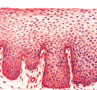
The images below show a magnified cross section of the lining of the oral cavity and the throat. On the left is a normal example, and on the right is an example of squamous cell carcinoma. You can that the cancer is much darker in its coloration (the biopsy specimens are all colored with special stains that make identification easier). The cancer also shows invasion into the underlying tissue layers, while the normal tissue has an orderly appearance. The pathologists who make the diagnosis rely on these and many other clues to distinguish normal tissue from cancerous growths.
 |
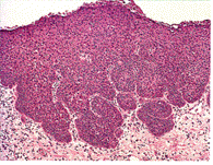 |
| Normal cells | Squamous cell carcinoma |