behind The Bench
EVMS researchers extract new data from prostate cancer biopsies
Researchers at EVMS say they have discovered a new way to preserve and measure tissue that allows for additional testing from a single biopsy.
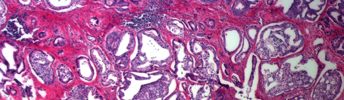
In a typical biopsy, a physician obtains a tissue sample and sends it to a laboratory for analysis. It may be chemically treated or frozen by a pathologist, then cut into sections to be examined under a microscope. Once the tissue has been altered for screening under the microscope, it can’t be tested for metabolites.
In a collaboration with the University of Alberta, Canada, researchers discovered that if the tissue is preserved in alcohol, instead of the more commonly used formalin, the preserving liquid also can be tested for metabolites.
The method is called molecular preservation by extraction and fixation (mPREF). In a proof-of-principle study, researchers used the method to study 25 samples from patients undergoing a prostatectomy. They found that 2,900 metabolites were consistently detected in more than 50 percent of the samples. This unprecedented coverage helped identify significant metabolites for differentiating tumor and normal tissues.
“Our long-term goal of using mPREF in prostate cancer is to identify metabolite biomarkers that are prognostic,” says Dean Troyer, MD, Professor of Microbiology and Molecular Cell Biology, Associate Director of Translational Research at the EVMS Leroy T. Canoles Jr. Cancer Research Center and co-lead author of the study. “This method offers a powerful and convenient means of performing histopathology and discovering or detecting metabolite biomarkers in the same tissue biopsy.”
While the results are promising, Dr. Troyer says the findings are preliminary because of a limited number of samples analyzed. Future work is needed to validate the prediction capability of these potential biomarkers using larger cohorts, ultimately including samples from multiple centers.
“Molecular pathology is a cornerstone to individualized medicine,” says John Semmes, PhD, Anthem Distinguished Professor for Cancer Research, Director of the Canoles cancer center and Professor of Microbiology and Molecular Cell Biology. “If realized within a viable clinical workflow, it provides companion diagnostics for improved patient care. Dr. Troyer’s discovery is a significant step toward bridging gold-standard pathology with discrete biopsy-derived molecular insight.”
In the future, Dr. Troyer believes the method will also be useful in toxicology, biopsies of transplanted organs and other cancers.
The study, “Metabolite Analysis and Histology on the Exact Same Tissue: Comprehensive Metabolomic Profiling and Metabolic Classification of Prostate Cancer,” was published in the journal Nature Scientific Reports.