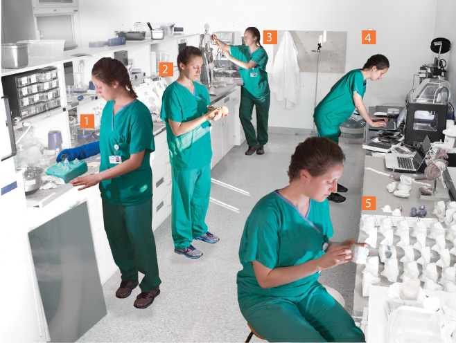

Follow Pathology and Anatomy Intern Katie Van Winkle as she constructs anatomically accurate models in her Lewis Hall lab. Medical and Health Professions students use her creations to better understand human anatomy.
- Katie pours hot ballistic gel into an eye mold to create normal and pathological eyes used in ultrasound training.
- She sculpts a clay mold for a 20-week fetus. The mold is filled with gel around an embedded, 3D-printed plastic skeleton and is used for ultrasound training.
- She uses clay to sculpt and position musculature on a three-foot, pre-made skeleton. The clay muscles help complete an atlas for students learning anatomy.
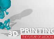
- She prepares the 3D printer. The printer uses melted plastic to create a model based on a computer-generated diagram.
- She places eyes crafted in stage 1 into a sculpted partial skull. These "ocular pathology trainers" are used.