Dissecting Contemporary Human Anatomy
A new twist on an old art
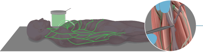
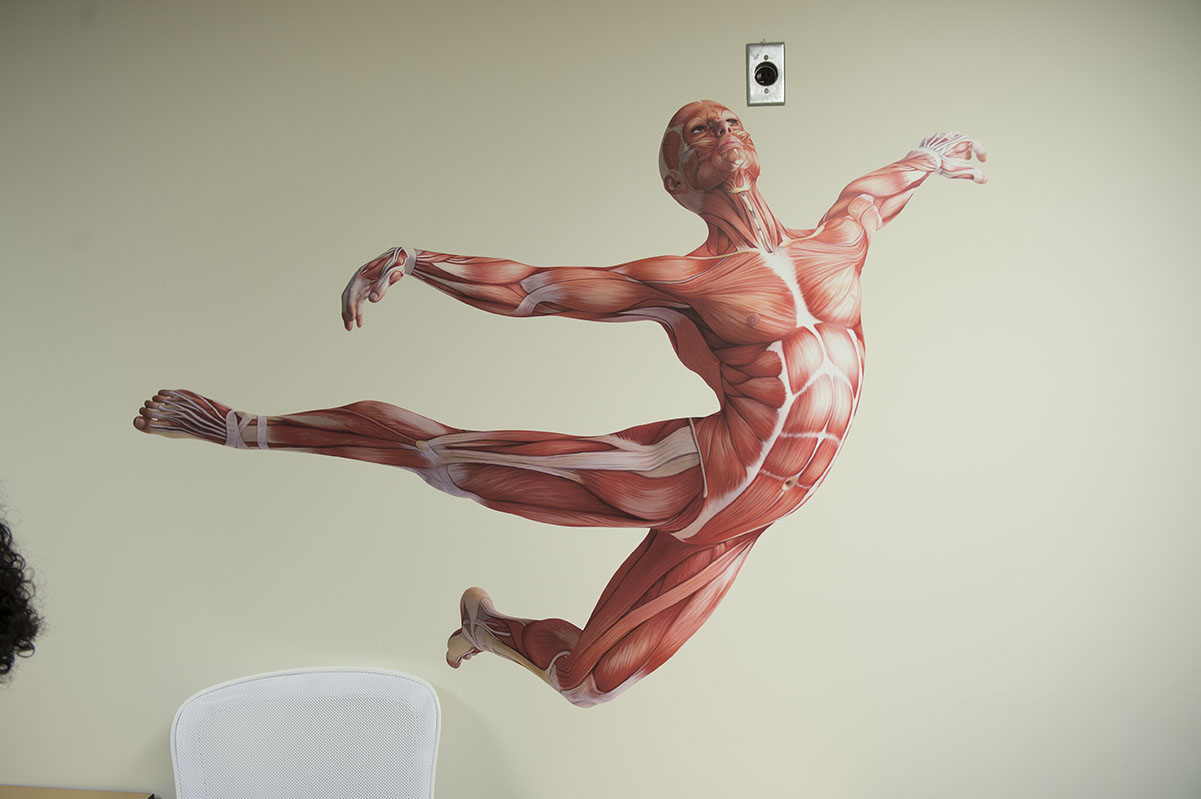
The boundaries between life and death — between skin and bone — are explored here.
A profusion of blood vessels extends over and through fleshy, pink tissue like rivers and tributaries across a dense landscape. Bones meet joints and ligaments where strings of muscle seemingly connect and overlap.
Carrie Elzie, PhD, stands silently at the front of the lab and observes the artistry of anatomy.
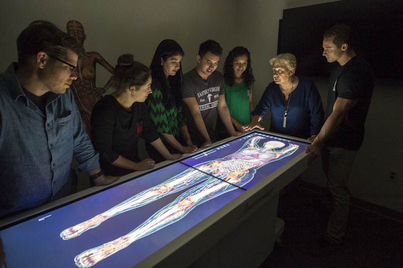
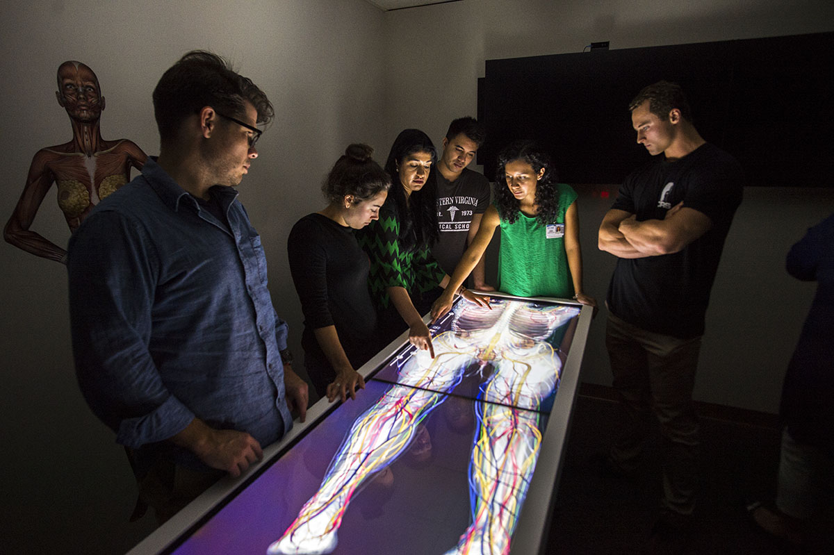
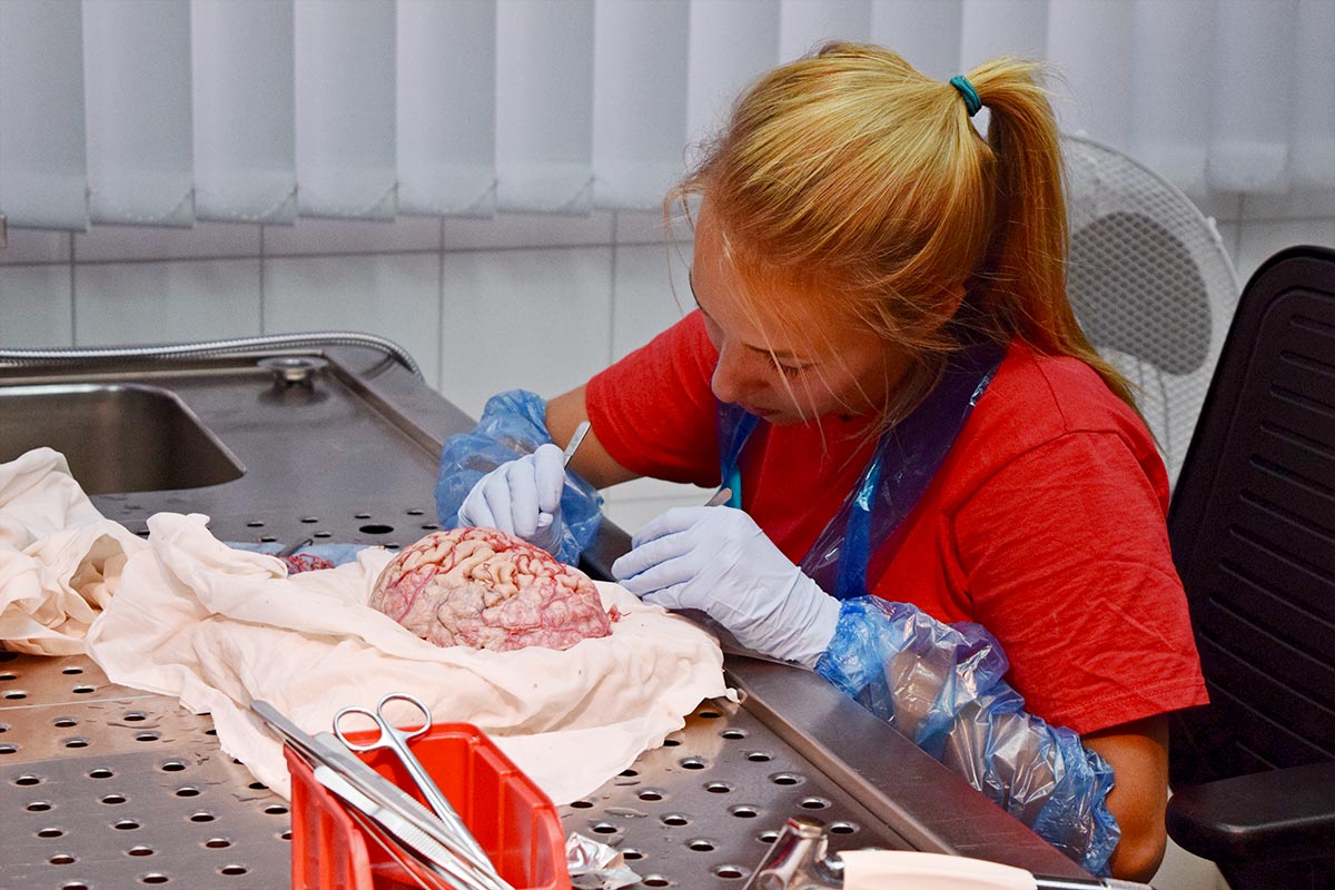
Today, her students are engrossed in full dissection. In pairs, they talk softly as they slice. On a different day, they will use imaging to turn, rotate and cut into cross-sections of a virtual cadaver with simple strokes of the hand.
Here, students are studying the complex systems of human anatomy in a true dichotomy of an age-old art and contemporary science.
“Medicine wasn’t my calling. I don’t have the knack for working on anybody who’s still breathing and whose life is in my hands,” says Dr. Elzie, Assistant Professor of Pathology and Anatomy. “But I certainly have the fascination of understanding the intricacies of the human body and the disease processes it can take on.”
What Dr. Elzie does have a knack for is teaching students and helping them discover a career path. That knack, plus her passion for anatomy, is what led her to launch the new Master of Contemporary Human Anatomy (CHA) program at EVMS.
It’s called ‘contemporary’ because it’s learning the human body through medical imaging and dissection,” she says, “so it’s viewing the body differently than what we have been doing over the past 100 years in anatomy.”
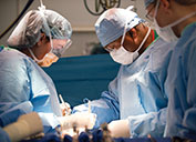
The three-semester program marries traditional sciences and classic anatomy coursework with emerging technology like virtual dissection, 3D printing and plastination. It begins in the summer with an intense seven-week gross-anatomy experience where students conduct a full dissection of a cadaver — inch by inch.
“I love the human body. I think it is the most amazing and complex system,” says Katie Van Winkle, CHA Class of 2017. “I love that a lot of it doesn’t make sense still — the whole intricacy of everything that is working in your body, and how every body that you dissect is different, and yet every one is still the same.”
Anatomy of the program
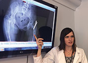
In their first semester, CHA students are taught gross anatomy and embryology, are introduced to the school’s renowned ultrasound curriculum and learn to read and interpret MRI and CT imaging.
By the fall, they work as teaching assistants in the anatomy classes of medical students and those in the Medical Master’s program. They take histology with medical students and a course in research in medical and health-professions education.
And because CHA is built to be a flexible master’s degree, students select the electives and capstone project during their spring semester that will best prepare them for their post-graduate goals.
The 10 students in the inaugural class have diverse career interests that include medical school, physician-assistant program, medical illustration, anatomy education and medical-model engineering.
For Malbika Ramchandani, CHA Class of 2017, the CHA program is a first step toward medical school. That’s been the lifelong dream of the Virginia Beach resident who attended the Health and Sciences Academy at Bayside High School.
“I remember being 14 and Dr. Goodmurphy visiting our class with his box of organs to teach us about lungs and kidneys,” Ms. Ramchandani says. “That made me choose medicine.”
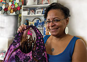
This spring, Ms. Ranchandani will spend a portion of the semester at the Plastinarium in Guben, Germany, where she will learn the science of the body-preservation techniques of plastination from its creator, Gunther von Hagens.
It will be yet another way for Ms. Ranchandani to explore human anatomy and the impact of the field of medicine.
“Medicine is evolving, and you have to keep growing with it,” she says. “This program makes me want to push myself.”
From scalpel to screen
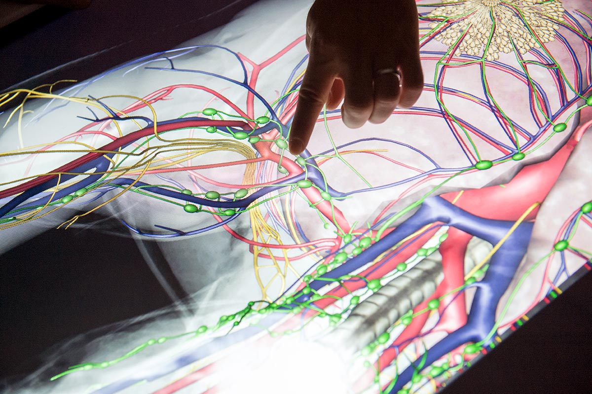
Tucked in a classroom in the back corner of Lewis Hall is a virtual dissection table — think of a table-sized iPad — that enables CHA students to practice dissection techniques when they can’t work in the cadaver lab. It’s yet another way that CHA sets itself apart from traditional anatomy programs.
On the opposite hall, CHA students work in the school’s 3D-printing lab, creating medical simulators that will be used by medical and health-professions students alike.
Like other contemporary anatomy programs across the country, EVMS has introduced multimodal learning in the lab.
Rick Drake, PhD, an anatomist for 40 years, has written the textbook used by many schools and universities today. He is currently Director of Anatomy at the Cleveland Clinic Lerner College of Medicine and serves as the Co-Editor-in-Chief of the Anatomical Sciences Education Journal, which publishes much on the topic of anatomy education and training.
He considers the addition of technology in the anatomy lab as a contemporary approach to an old art.
“Anatomy is a very visual subject that’s difficult to get your head around without the benefit of dissection,” Dr. Drake says. “Technology has its place. What is important is to figure out how it works best and to use it where it has the biggest impact.”
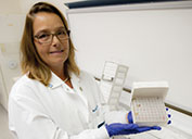
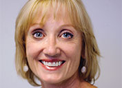
Rebecca Lufler, PhD, Chair of the Educational Affairs Committee of the American Association of Anatomists, says the continuous growth in medical technology and the introduction of programs like CHA highlights a need in the field.
“I think there is a great need for teachers of anatomy in all programs — from allied health to medical school to undergraduate,” she says. “The more anatomists we can train, the better.”
Especially since the field is quickly changing, she says.
“Medicine is evolving, and you have to keep growing with it,” she says. “This program makes me want to push myself.”
A classically trained anatomist, Dr. Lufler now teaches anatomy at Tufts University School of Medicine in a high-tech lab that integrates radiology with traditional anatomy.
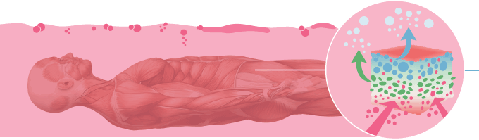
From textbooks and apps to web-based learning and imaging, human anatomy courses have taken a big step into the technological age.
“Using technology to assist traditional anatomy only enhances the experience,” Dr. Elzie says. “It allows our students to learn things in a more three-dimensional aspect and have a little bit more control over that.”
That integration of technology is a centerpiece of the CHA curriculum — not to replace cadaver dissection, but to complement it.
"The actual experience of dissection is not replaceable,” Dr. Elzie says. “Instead, this is a whole new level of learning that targets the millennial learner.”
Learners like Aaron Magana, CHA Class of 2017, who was surprised at the diverse opportunities provided by the CHA program.
An aspiring future doctor, Mr. Magana was recently accepted to an osteopathic medical school and will begin classes shortly after graduating from EVMS.
“I don’t think I could have gotten that far without this program,” he says.
Fellow CHA classmate Jonathan Krimsier agrees.
“If you like medicine, but don’t want to practice it, this is a great place to start because it will expose you to all the other avenues involved with medicine, be it education or anything else behind the scenes,” says Mr. Krimsier, who hasn’t yet decided where his master’s degree will take him next. “I just know this is the first step on my road.”